KNEE OCD
Living with Osteochondritis Dissecans (OCD) of the knee can feel overwhelming, whether you are a patient experiencing symptoms or a parent trying to guide your child through the process. The uncertainty around diagnosis, treatment options, and recovery often raises many questions. This section is designed to bring clarity and reassurance by offering information that is straightforward, accurate, and easy to follow.
Here you will find explanations of how OCD is diagnosed, what treatments may be recommended, and what recovery typically looks like. Each topic is broken down into smaller steps, supported by visuals that make the information easier to understand. Our goal is to help patients and families feel informed and supported, not only in understanding the medical facts but also in preparing for the day-to-day challenges of living with and managing OCD of the knee.
By making complex information easier to digest, we hope this resource empowers you to have meaningful conversations with your care team, make confident decisions, and feel more at ease about the road ahead.

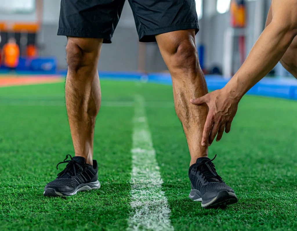
Pain is usually the first sign of Osteochondritis Dissecans. It often worsens with physical activity and improves with rest. Swelling may develop around the joint, especially after exercise, and can make the knee feel tight or heavy.


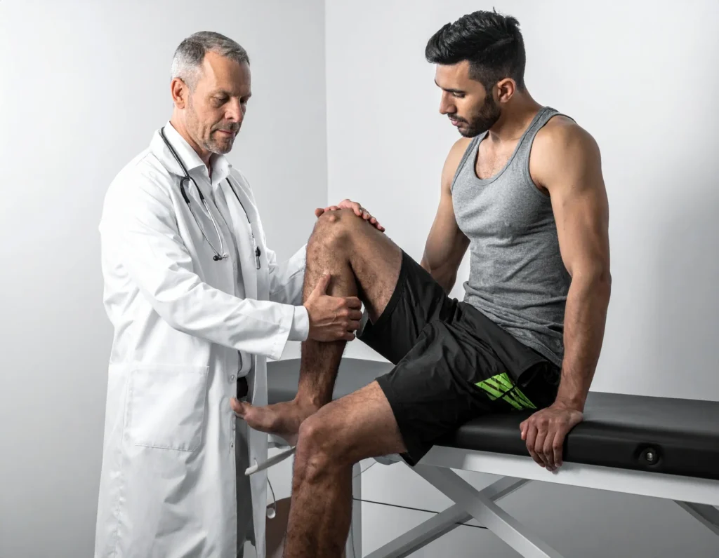
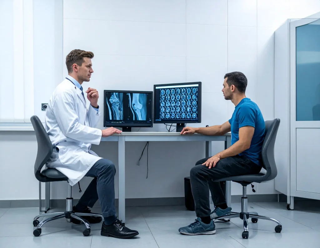
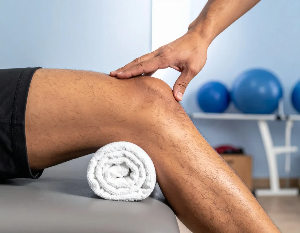
Diagnosing Osteochondritis Dissecans (OCD) of the knee usually begins with a careful physical exam. Your doctor will check for tenderness, swelling, limited range of motion, and may ask you to describe your pain during activity or rest. Because OCD affects both the bone and cartilage, imaging tests are an important part of confirming the diagnosis. Standard X-rays give a clear view of the bone structure and can reveal changes in the shape or density of the affected area. In many cases, an MRI is also ordered to provide a more detailed picture, showing not only the bone but also the overlying cartilage and the stability of the fragment. Together, these steps allow doctors to determine how advanced the condition is and guide the choice of treatment that will best protect the knee and support long-term healing.
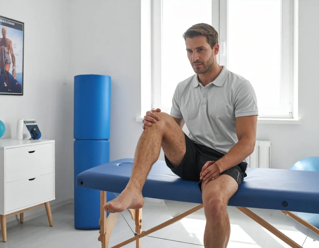
During the physical exam, your doctor will look closely at how your knee moves and feels. They may press on different parts of the joint to check for tenderness or swelling, and gently move the knee through its range of motion to see if it causes pain or stiffness. You might be asked to walk, bend, or perform simple movements so the doctor can observe how your knee responds under pressure. These tests help identify where the problem is located and whether OCD may be the cause of your symptoms.
X-rays are often the first imaging test used to evaluate OCD of the knee. They provide a clear picture of the bone and can show changes in shape, density, or small areas where the bone may have weakened. In some cases, X-rays can also reveal whether a fragment of bone and cartilage has started to separate. Doctors typically take images from different angles to get the most accurate view. While X-rays cannot show cartilage, they are a quick and reliable way to confirm that OCD may be present and to track changes over time.

An MRI provides the most detailed view of Osteochondritis Dissecans (OCD) of the knee. Unlike X-rays, which only show bone, an MRI reveals both the bone and the surrounding cartilage. This allows doctors to see whether the OCD lesion is stable or if a fragment has loosened. MRI scans can also show early changes in the cartilage before they appear on an X-ray, making them especially helpful in younger patients or in cases where the condition is just beginning. The information from an MRI helps guide treatment by showing exactly how much of the knee is involved and whether surgery may be needed.
Treatment for Osteochondritis Dissecans (OCD) of the knee depends on the patient’s age, the size of the lesion, and whether the bone and cartilage are stable. In younger patients whose bones are still growing, rest, bracing, and careful activity modification often give the lesion time to heal naturally. Physical therapy may be added to restore strength, flexibility, and balance as the knee recovers. If the lesion is unstable or healing does not occur with non-surgical care, surgery may be recommended to repair or restore the joint. The overall goal of treatment is to protect the knee, relieve pain, and ensure the best possible long-term outcome for activity and sport.
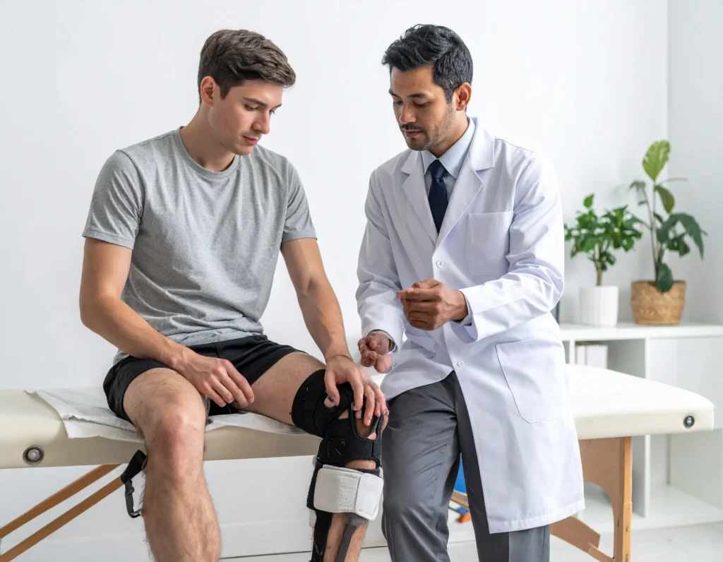
For many younger patients, the first step in treating Osteochondritis Dissecans (OCD) of the knee is giving the joint time to heal. Doctors often recommend a period of rest from high-impact activities like running and jumping, along with the use of a supportive knee brace. The brace helps limit stress on the affected area while still allowing safe, gentle movement. This approach gives the bone and cartilage the best chance to repair themselves naturally. While it can be challenging for active patients to take a break from sports, rest is one of the most effective tools for recovery, and it helps prevent the lesion from getting worse.
Physical therapy is a key part of recovery for patients with Osteochondritis Dissecans (OCD) of the knee. Once pain and swelling are under control, a therapist guides patients through exercises that gently restore strength, flexibility, and balance. Early therapy may focus on maintaining motion and preventing stiffness, while later stages emphasize building muscle support around the knee to protect the joint during daily activities and sports. A tailored program not only speeds up recovery but also helps reduce the risk of future injuries by improving overall knee stability.

When non-surgical treatments are not enough, surgery may be recommended to stabilize or repair the lesion caused by Osteochondritis Dissecans (OCD) of the knee. The specific procedure depends on the size and stability of the fragment. In some cases, small holes are made in the bone to stimulate healing, while in others, the loose piece of bone and cartilage is secured with small pins or screws. If the fragment cannot be saved, a graft may be used to restore the joint surface. These procedures are performed by orthopedic surgeons who specialize in treating knee injuries, with the goal of preserving as much natural bone and cartilage as possible and supporting long-term knee health.

Recovery from Osteochondritis Dissecans (OCD) of the knee is a gradual process that varies for each patient. After treatment the main focus is on regaining motion, building strength, and restoring confidence in the knee. Physical therapy plays a central role, guiding patients through safe exercises that protect the healing joint while gradually increasing activity. Recovery also requires patience, as returning to sports or high impact activity too soon can put the knee at risk. With the right combination of rest, therapy, and follow up care most patients are able to return to the activities they enjoy, often with a stronger awareness of how to protect their knees in the future.
Gentle stretching is an important part of recovery from Osteochondritis Dissecans of the knee. Early in the process stretching helps maintain flexibility and prevent stiffness while the joint heals. Over time these exercises improve range of motion and make it easier to return to daily activities without discomfort. A physical therapist will guide which stretches are safe and how often they should be done, making sure the knee is supported and not pushed too quickly.

Returning to sports after treatment for Osteochondritis Dissecans takes time and careful planning. Before resuming full activity the knee must regain strength, stability, and confidence. Doctors and physical therapists use progress checks to decide when it is safe to advance. The process often begins with low impact exercises, then light sport specific drills, and finally full participation once the knee responds without pain or swelling. Following this step by step approach helps prevent re injury and ensures a safe return to the activities patients enjoy.
Learn about the doctors and centers dedicated to advancing Knee OCD care worldwide.
Get clear information on what it is, who it affects, and how it’s diagnosed.
Explore physical therapy routines and strategies that support healing and long-term joint health.
Access educational articles, guides, and tools designed to help you and your family navigate care.